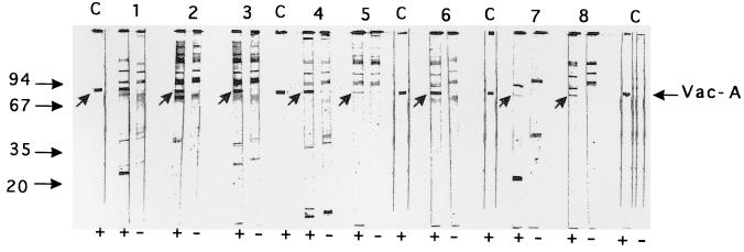
FIG. 2.
Immunoblot patterns obtained with vacuolating toxin-enriched preparations from the VacA-positive strain 60190 (+) and the isogenic VacA-negative mutant 60190:v1 (−) with eight selected patients’ sera which reacted with p87 from H. pylori ATCC 43579 (lanes 1 to 8). Lanes C, serum from a rabbit immunized to purified VacA. The arrows indicate immunoreactive bands corresponding to VacA. Molecular mass markers are on the left. The figure shows a scan of the original nitrocellulose strips.

