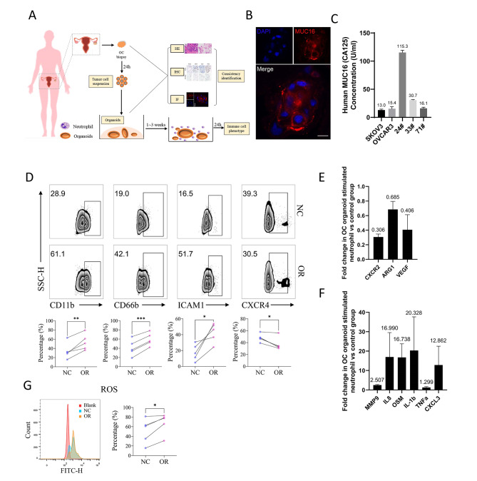
Fig. 2.
Patient-derived ovarian cancer organoids altered the immune phenotype of neutrophils. (A) Flow chart for the establishment of patient-derived ovarian cancer organoids. (B) Expression of MUC16 in ovarian cancer organoids (24#) determined by immunofluorescence under the confocal microscope, bar = 10 μm. (C) MUC16 level in the supernatants of ovarian cancer cell lines and ovarian cancer organoids detected by ELISA assay. (D) The proportion of CD11b+, CD66b+, ICAM-1+, CXCR4+ neutrophils in the ovarian cancer organoid stimulation group (OR) and control group (NC). The upper panel shows the representative flow cytometry results; the lower panel shows the experimental results of 5 cases of neutrophils from different patients. Paired t-test. (E-F) The expression of function-related factors in the ovarian cancer organoid stimulation group compared to the control group determined by qPCR (three independent replicate experiments of neutrophils derived from different patients). Error Bar = Mean ± SEM. (G) ROS detection of ovarian cancer organoid stimulation group (OR) and control group (NC). Representative results were shown on the left. Blank: blank control group without DCFH-DA; NC: negative control group without ovarian cancer organoid stimulation; OR: ovarian cancer organoid stimulation group. N = 5, paired t-test. (*p < 0.05, **p < 0.01, ***p < 0.001, ****p < 0.0001.)

