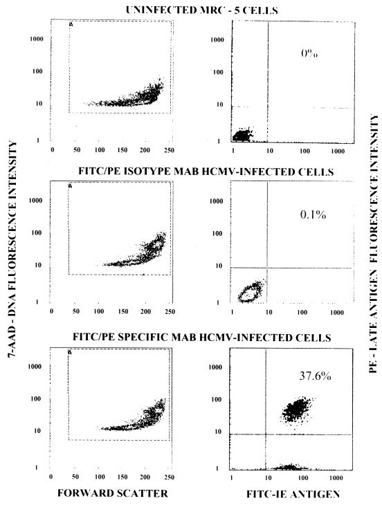
FIG. 1.
Three-color flow cytometric analysis of uninfected and HCMV-infected cells. Left-hand panels display results with uninfected and HCMV-infected MRC-5 cells analyzed for DNA content (7-AAD) versus cell size (forward scatter). Right-hand panels display results with uninfected and HCMV-infected cells analyzed for late-antigen-positive cells versus immediate-early (IE)-antigen-positive cells. MAB, monoclonal antibody.

