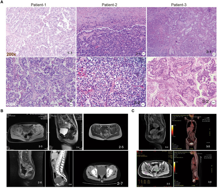
Figure 1.
Pathologic examination of juvenile CCAC independent of HPV. (A) Patient-1, papillary growth areas with relatively small and regular papillae, sometimes larger papillae, with fibrovascular axis, tumor composed of moderately to severely atypical cells with clear cytoplasm and polygonal cells; patient-2, the tumor was composed of moderately to severely atypical cells with clear cytoplasm and polygonal cells; patient-3, in the papillary growth area, the papilla are small and relatively regular, sometimes larger, with fibrovascular axis, “shoe spike” cells, and cells protruding into the lumen of the gland. Scale: top row: 200×, bottom row: 400×. (B) Pelvic MRI images of patient-2 at initial diagnostic stages (2-3, 2-4); suggestive of recurrence stages (2-5, 2-6); and follow-up CT images showing no signs of recurrence (2-7, 2-8). (C) Pelvic MRI images of patient-3 at initial diagnosis (3-3, 3-4); follow-up PET-CT images showed no signs of recurrence (3-5, 3-6).

