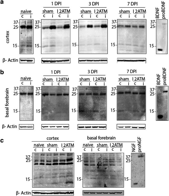
Figure 1.
Proneurotrophins are induced in the ipsilateral cortex but not the basal forebrain after cortical FPI. a–c, Brain tissue lysates from naive, sham, and injured (2 atm) wild-type adult mice were obtained 1DPI, 3DPI, and 7DPI to determine levels of proBDNF (a, b) and proNGF (c) in the injured versus uninjured side. Cortical tissue lysate (a) harvested for Western blot was probed for proBDNF (32 kDa) in the ipsilateral and contralateral cortex at 1DPI, 3DPI, and 7DPI in naive, sham, and injured mice. Basal forebrain tissue lysate (b) harvested for Western blot was probed for proBDNF (32 kDa) in the ipsilateral versus contralateral basal forebrain at 1DPI, 3DPI, and 7DPI in naive, sham, and injured mice. Cortex and basal forebrain tissue lysates (c) harvested for Western blot were probed for proNGF (37 kDa) at 3DPI after FPI in the ipsilateral versus contralateral side of the cortex and the basal forebrain; n = 4 (naive), n = 4 (sham 1DPI), n = 4 (injured 1DPI), n = 4 (sham 3DPI), n = 4 (injured 3DPI), n = 3 (sham 7DPI), n = 3 (injured 7DPI; a, b); n = 3 (naive), n = 4 (sham 3DPI), n = 4 (injured 3DPI; c). The established size of proBDNF is 32 kDa; however, a prominent band of 25 kDa was also recognized by the BDNF antibody that appeared to be regulated by injury, but the identity of that band is unclear.

