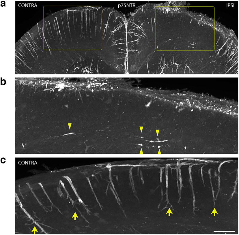
Figure 5.
Cortical FPI leads to a retrograde axonal degeneration of afferent basal forebrain neurons ipsilateral to the injury 7DPI. a–c, WT mouse brains were fixed 7DPI after cortical FPIs were cleared by iDISCO before immunostaining for p75NTR. Whole brains were analyzed by light sheet microscopy. Areas highlighted in rectangles (yellow) are magnified (b, c) to show the IPSI and CONTRA regions in further detail. p75NTR staining ipsilateral to the injury (b) shows p75NTR+ basal forebrain afferents with varicosities, tortuosity, and retraction bulbs (yellow arrowheads). p75NTR staining in the cortex contralateral to the injury (c). Yellow arrows denote p75NTR+ blood vessels in the uninjured cortex; n = 3 (7DPI). Scale bar, 50 μm.

