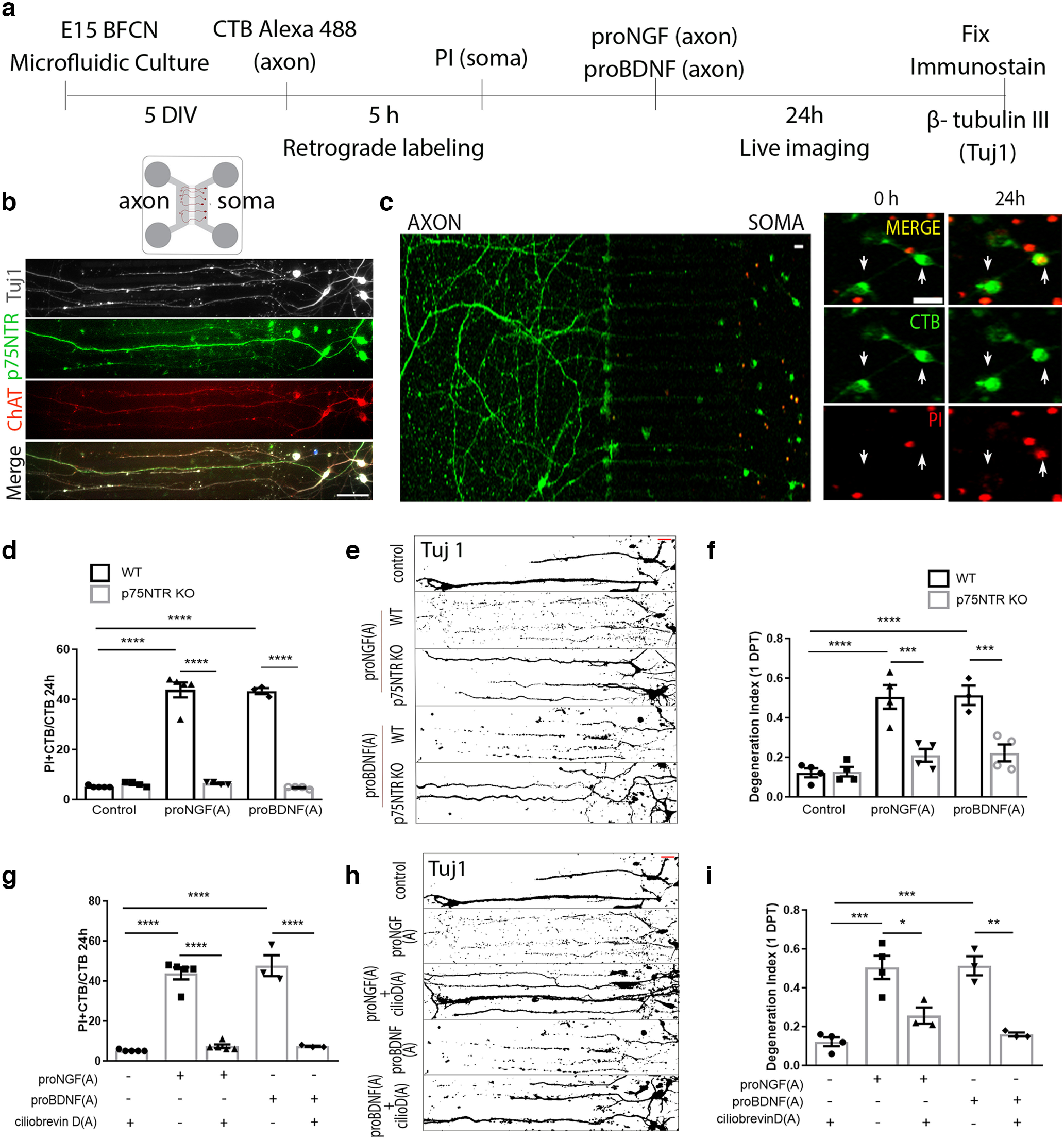
Figure 6.

proNGF and proBDNF promote retrograde degeneration of BFCNs in microfluidic cultures via p75NTR. a, Basal forebrain neurons were cultured from E15 mouse embryos in microfluidic chambers for 5DIV. b, BFCNs grown in microfluidic chambers coimmunolabeled for Tuj1 (gray), p75NTR (green), and ChAT (red). Scale bar, 50 μm. c, The axon compartment was treated with CTB Alexa 488 (green) to retrogradely trace neurons that extended their axons to the distal compartment. PI (red) was added to the soma compartment before axonal treatment to study dying (PI+/CTB+) neurons after 24 h of axonal treatment with proneurotrophins. Arrows indicate CTB+ neurons that incorporate PI in their nucleus after 24 h of treatment. Scale bar, 20 μm. d, Quantification of dying neurons in WT and p75NTR KO cultured BFCNs after axonal treatment with proNGF or proBDNF; n = 5 (Control, WT), n = 4 (Control, KO), n = 5 [proNGF(A), WT)], n = 4 [proNGF(A), KO], n = 3 [proBDNF(A), WT], n = 4 [proBDNF(A), KO], where (A) indicates axonal treatment; ****p < 0.0001 by two-way ANOVA, Sidak’s multiple comparisons tests. e, Axon fragmentation in WT or KO BFCNs after proNGF or proBDNF treatment assessed using Tuj1 staining represented as binary images. Scale bar (red, top right), 20 μm. f, Quantification of axonal degeneration in WT and p75NTR KO cultured BFCNs after axonal treatment with proNGF or proBDNF; n = 4 (Control, WT), n = 4 (Control, KO), n = 4 [proNGF(A), WT], n = 4 [proNGF(A), KO], n = 3 [proBDNF(A), WT], n = 4 [proBDNF(A), KO]; ****p < 0.0001 comparing WT/control versus proNGF(A), and WT:control versus proBDNF(A), ****p = 0.0007 comparing WT:proNGF(A) versus KO:proNGF(A), ***p = 0.0007, and WT:proBDNF(A) versus KO:proBDNF(A) ***p = 0.0007 by two-way ANOVA, Sidak’s multiple comparisons tests. g, Quantification of dying neurons in WT cultured BFCNs with or without pretreatment with ciliobrevin D (50 μm) before proNGF or proBDNF treatment in the axons; n = 5 (Control), n = 5 [proNGF(A)], n = 5 [proNGF(A)+ CilioD(A), KO], n = 3 [proBDNF(A)], n = 3 [proBDNF(A)+ CIlioD(A)]; ****p < 0.0001 by one-way ANOVA, Tukey’s multiple comparison tests. h, i, Axon fragmentation in WT cultured BFCNs with or without pretreatment with ciliobrevin D (50 μm) before proNGF or proBDNF treatment in the axons assessed using Tuj1 staining represented as binary images (h) and quantification of axonal degeneration (i); n = 4 (Control), n = 4 [proNGF(A)], n = 3 [proNGF(A)+ CilioD(A)], n = 3 [proBDNF(A)], n = 3 [proBDNF(A)+ CilioD(A)]. Scale bar, h (red, top right), 20 μm. In i the asterisk indicates ***p = 0.0001 comparing control versus proNGF(A), ***p = 0.0002 comparing control versus proBDNF(A), p = 0.0102 comparing proNGF(A) versus proNGF(A)+ CilioD(A), and **p = 0.0011 comparing proBDNF(A) versus proBDNF(A)+ CilioD(A) by one-way ANOVA, Tukey’s multiple comparison tests.
