
FIGURE 1.
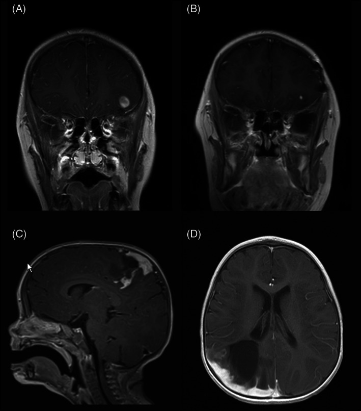
MRI findings in TRAK1::RAF1 fusion positive gliomas: T1‐weighted, post‐contrast MRI demonstrates a hypointense, well‐circumscribed 1 cm ovoid lesion with heterogenous rim‐like enhancement, cortically‐based in the left inferotemporal lobe in Case 1 (A); after gross total resection, an area of focal nodular enhancement subjacent to the surgical site was noted on 14‐month surveillance scan (B). Tumors in Case 2 (C) and Case 3 (D) involved both brain and leptomeninges. T1‐weighted, post‐contrast MRI in Case 2 demonstrated a 3 cm solid and cystic enhancing mass with significant surrounding edema (C), while T1‐weighted, post‐contrast MRI in Case 3 demonstrated an infiltrative lesion in the R parieto‐occipital lobe with thick nodular leptomeningeal enhancement and ~4 cm non‐enhancing cystic‐solid component (D).