
FIGURE 2.
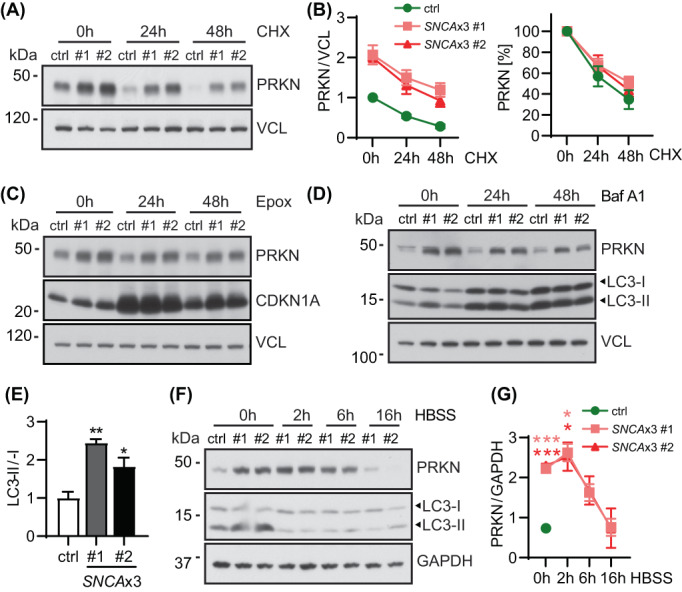
Activation of general autophagy reduces PRKN accumulation in SNCAx3 fibroblasts. (A) Representative western blot of control and SNCAx3 fibroblasts treated with cycloheximide (CHX) for the indicated timepoints. (B) Western blot quantification of PRKN normalized with the value of the control line set to 1 (left) or using each value at 0 h as 100% (right). Results show comparable PRKN turnover rates in all three fibroblast lines. n = 4 independent experiments. (C) Proteosome inhibition does not affect PRKN levels in control or SNCAx3 fibroblasts. Representative western blot of PRKN, CDKN1A, and VCL from control and SNCAx3 fibroblasts after 1 μM epoxomicin (Epox) treatment for 0, 24, and 48 h. (D) Inhibition of autophagic flux does not further increase PRKN levels in SNCAx3 fibroblasts. Representative western blot of PRKN, LC3, and VCL from control and SNCAx3 fibroblasts after 200 nM bafilomycin A1 (Baf A1) treatment for 0, 24, and 48 h. (E) Immunoblot quantification shows significant increases of lipidated to unlipidated LC3 ratio (LC3‐II/I) in two SNCAx3 fibroblasts compared to control (p = 0.0022 for #1, p = 0.031 for #2). Quantifications are shown as fold change with the control set to 1. n = 3 independent experiments. (F) Representative western blot of control and SNCAx3 fibroblasts treated by HBSS for indicated timepoints. (G) Western blot quantification shows that HBSS‐induced autophagy activation is able to lower PRKN levels in SNCAx3 fibroblasts to the control level after 16 h of starvation. n = 2 independent experiments. Data shown as mean with standard error. One‐way ANOVA, *p < 0.05, **p < 0.01, ***p < 0.001 in pink/red for the corresponding SNCAx3 cells when compared to control at the same timepoint.