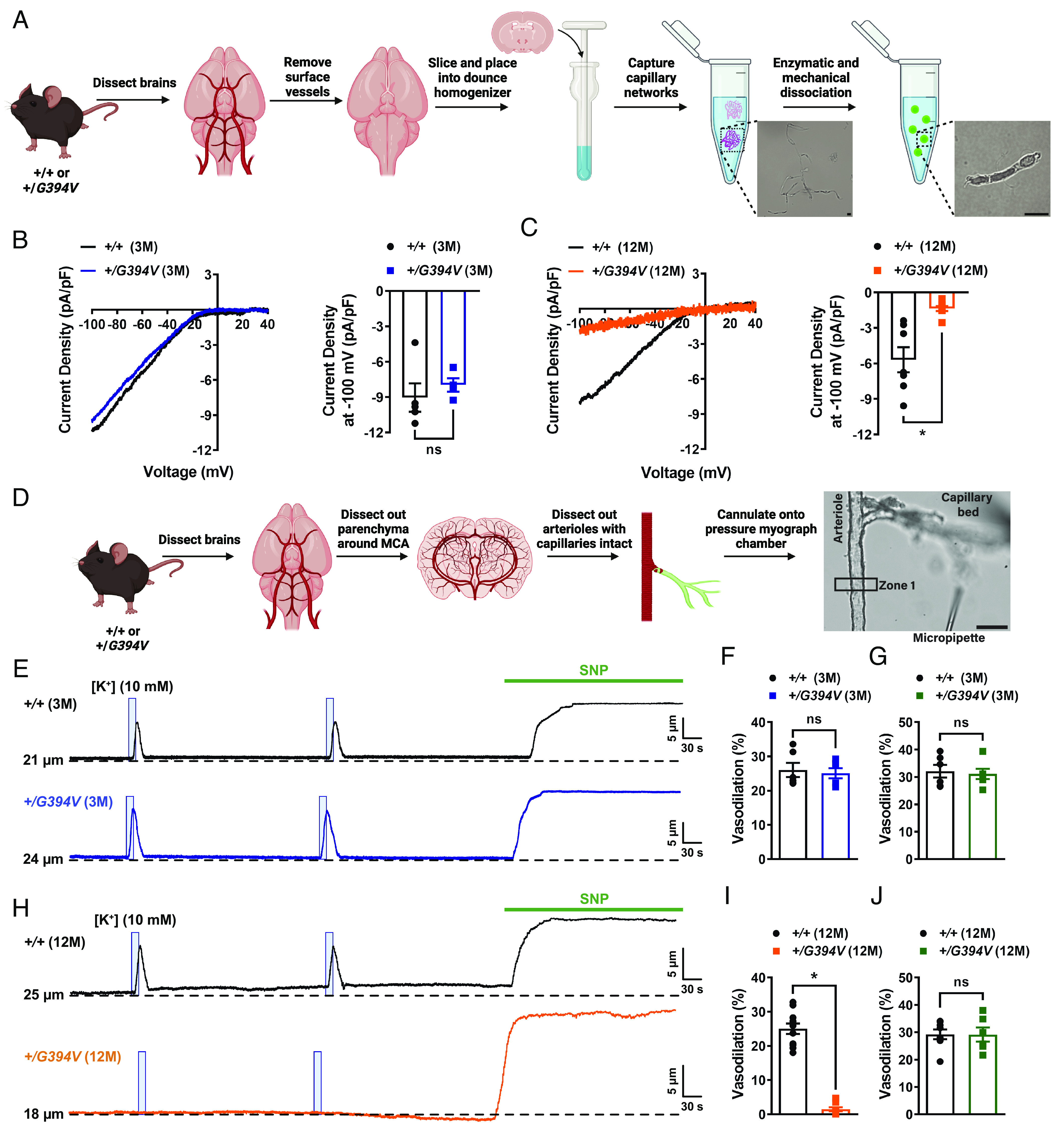
Fig. 1.

Age-dependent loss of Kir2.1 channel activity and capillary-to-arteriole dilation in Col4a1+/G394V mice. (A) Illustration of the brain capillary EC isolation procedure. (Scale bar, 10 µm.) (B) Representative I-V traces and summary data showing Kir2.1 current densities in freshly isolated capillary ECs from 3-M-old Col4a1+/+ and Col4a1+/G394V mice (n = 4 to 5 cells from 4 to 5 animals per group, ns = not significant, unpaired t test). (C) Representative I-V traces and summary data showing Kir2.1 current densities in freshly isolated capillary ECs from 12-M-old Col4a1+/+ and Col4a1+/G394V mice (n = 7 to 8 cells from four animals per group; *P < 0.05, unpaired t test). (D) Illustration of the microvascular preparation. Parenchymal arterioles with intact capillaries were carefully dissected and cannulated onto a pressure myograph chamber, and compounds of interest were focally applied to capillary extremities. (Scale bar, 50 µm.) (E and F) Representative traces (E) and summary data (F) showing K+ (10 mM, blue box)-induced dilation of upstream arterioles in preparations from 3-M-old Col4a1+/+ and Col4a1+/G394V mice (n = 6 preparations from three animals per group, ns = not significant, unpaired t-test). (G) The dilation produced by superfusing SNP (10 µM) in preparations from 3-M-old Col4a1+/+ and Col4a1+/G394V mice (n = 6 preparations from three animals per group, ns = not significant, unpaired t-test). (H and I) Representative traces (H) and summary data (I) showing K+ (10 mM, blue box)-induced dilation of upstream arterioles in preparations from 12-M-old Col4a1+/+ and Col4a1+/G394V mice (n = 11 preparations from 6 to 7 animals per group, *P < 0.05, unpaired t-test). (J) The dilation produced by superfusing SNP (10 µM) in preparations from 12-M-old Col4a1+/+ and Col4a1+/G394V mice (n = 6 to 8 preparations from 3 to 5 animals per group, ns = not significant, unpaired t-test).
