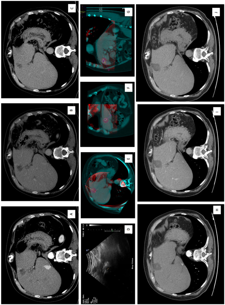
Figure 4.
150 W microwave ablation (MWA) of residual hepatic lesion after loco-regional treatment in S4a. (A) Arterial, (B) venous, (C) delayed phase show CECT show a contrast enhanced residual liver lesion with wash-out in venous and in delayed phases. (D) Intra-procedural US guidance during the insertion of MWA antenna. (E) Axial, (F) coronal, (G) sagittal plans imaging-fusion between intra-procedural CBCT and previous CECT, using navigational softwares, Xperguide (Philips Allura Xper FD20; Philips Healthcare, Best, Netherlands) for needle trajectory planning, and XperCT (Philips Allura Xper FD20; Philips Healthcare, Best, Netherlands), for the ablation volume prediction. (H) Arterial, (I) venous, and (J) delayed phases at 1-month CT follow-up, showing a complete response with the evidence of the coagulation zone. Abbreviations: MWA, microwave ablation; CBCT, cone-beam CT; CECT, contrast-enhanced CT; HCC hepatocellular carcinoma.

