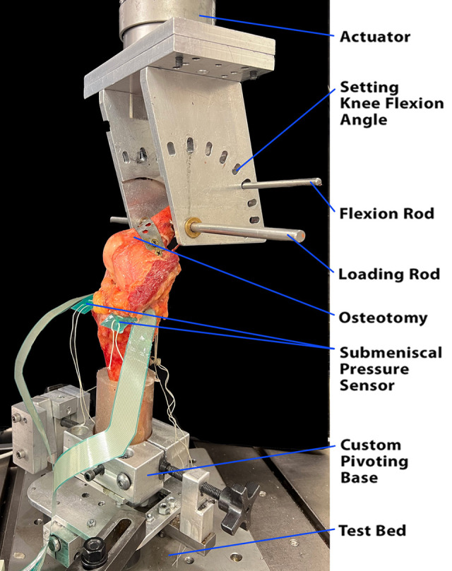
Figure 1.

The right cadaveric knee was held at 30° of flexion and loaded in a dynamic tensile machine. Each specimen underwent an oblique medial femoral condyle osteotomy to facilitate access to the medial compartment. The osteotomy was secured with a removable steel plate and bicortical screws. A transepicondylar “loading rod” (10-mm diameter) was placed medial to lateral and acted as the loadbearing axis during testing. An additional “flexion rod” (8-mm diameter) was passed medial to lateral through the proximal femur and allowed for changes to the knee flexion angle from 0° to 90° of flexion in 30° increments. The potted distal tibia was rigidly fixed to a custom pivoting base that allowed for freedom of motion in the transverse plane and for adjustment of the tibial orientation to standardize varus and valgus positioning.
