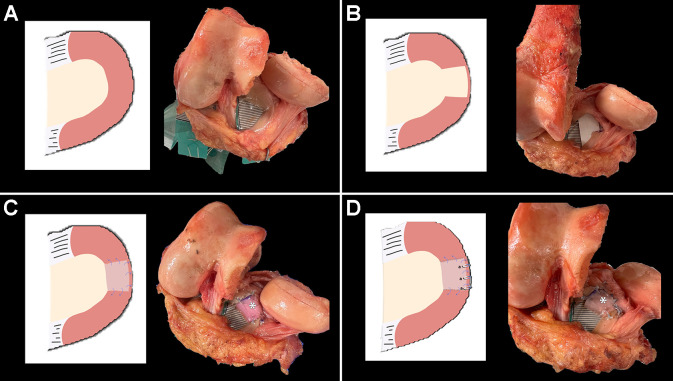
Figure 2.
Illustration and photograph of the medial meniscus of a right cadaveric knee in each testing state. (A) Intact state. (B) Segmental defect with a 15-mm tear of the midbody medial meniscus. (C) Inside-out segmental repair. A size-matched medial meniscal allograft was used on each specimen and secured in the anatomic position. Four sutures were passed at the anterior and posterior margins (2 in each margin) of the graft and secured with the remnant of the native meniscus in a horizontal mattress fashion. Then, 3 sutures were passed at the peripheral portion of the graft and repaired to the capsule 4 mm apart in a vertical mattress fashion (inside-out–type repair). (D) Anchor plus inside-out segmental repair. Segmental meniscal allograft transplantation (MAT) was augmented with 3 intracapsular knotless suture anchors placed at the rim of the medial tibial plateau 4 mm apart from each anchor. *Segmental MAT.

