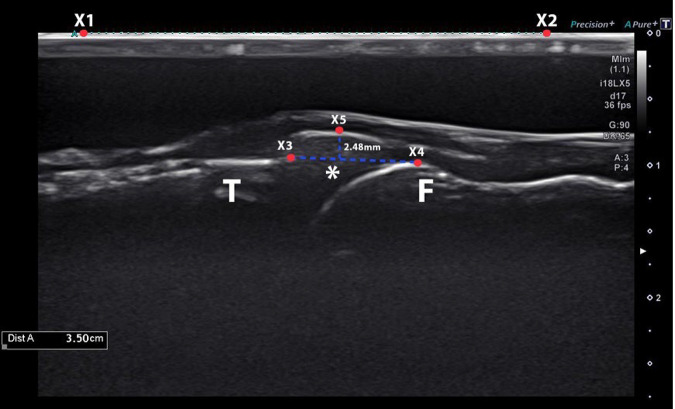
Figure 4.
An ultrasound image from a single specimen in the segmental defect state and at 60° of knee flexion. Medial meniscal extrusion was defined as the maximum distance between the medial margin of the meniscus (X5) and the line connecting the medial margin of the distal femur (X4) to the medial margin of the proximal tibia (X3), which was calibrated by the line measurement (X1 to X2) with the ultrasound machine. *Medial meniscus. F, distal femur; T, proximal tibia.

