Figure 2.
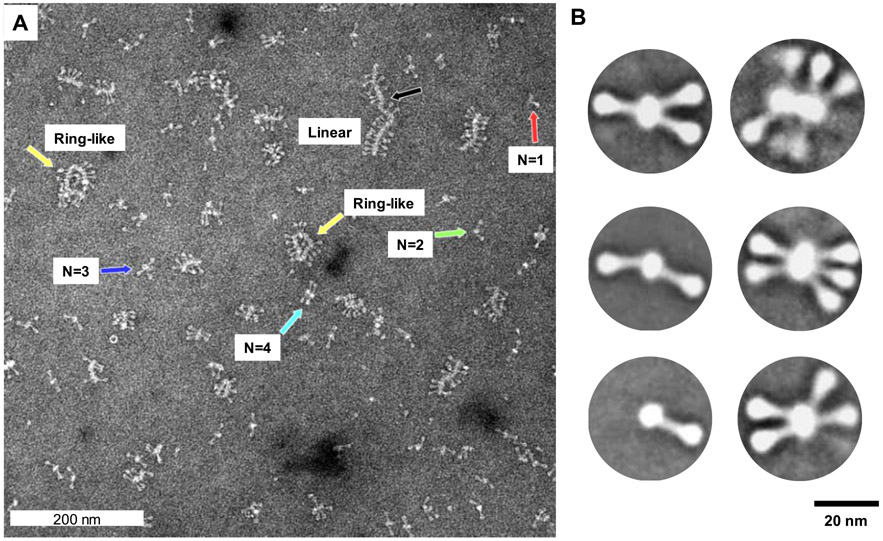
Transmission electron microscopy and 2D imaging of RSV F/PS80 nanoparticles. (A) Representative electron microscopy images of negatively stained RSV F/PS80 nanoparticles at a magnification of 52 000 × (0.21 nm/pixel). Discrete nanoparticles of a single RSV F trimer (red arrow), two RSV F trimers (green arrow), three RSV F trimers (blue arrow), four RSV F trimers (teal), large ring-like oligomers of 10–12 RSV F trimers (yellow arrows), and large oligomers of 14 or more RSV F trimers organized end-to-end (black arrow). (B) 2D class average images of nanoparticles with one to six RSV F trimers. The RSV F trimers have a 16–22 nm length with a head of approximately 5–6 nm that tapers to a 2–3 nm wide stalk associated with a central core of width approximately 8–10 nm and variable length depending on the number of RSV F trimers.

