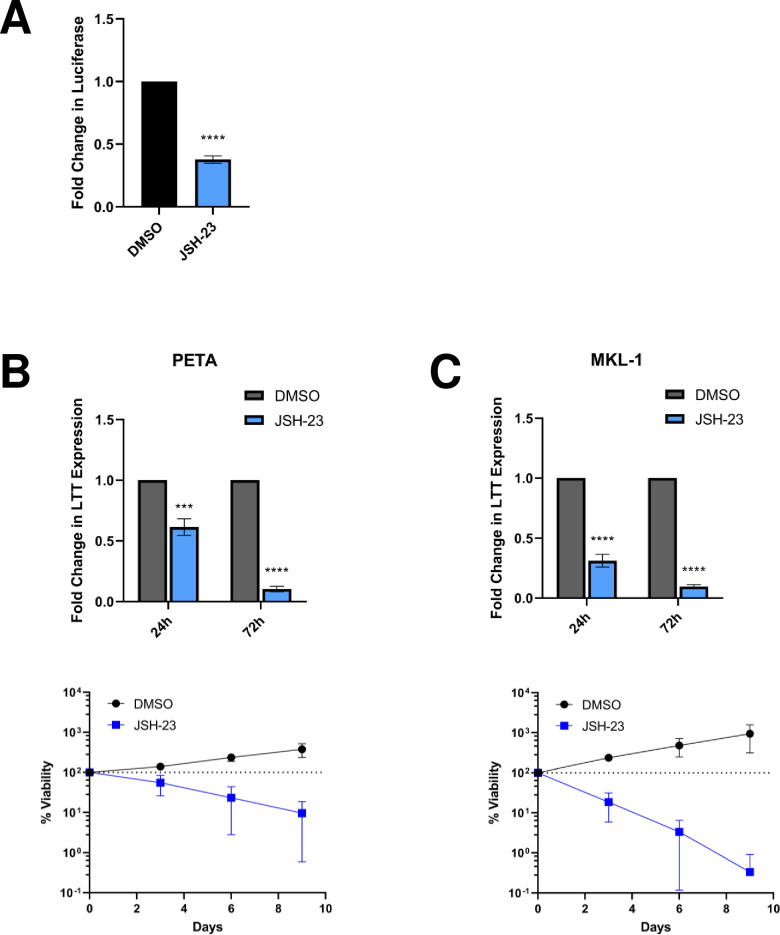
Fig 5. Inhibition of NF-κB activity represses MCPyV EP-driven viral oncogene expression, which is lethal in MCPyV+ MCC.
(A) HEK293 cells stably expressing an MCPyV EP-luciferase reporter were treated with DMSO or 25 μM JSH-23 for 72h before EP-driven luciferase expression was measured by luciferase assay. (B) PETA and (C) MKL-1 cells were treated with DMSO or 25 μM JSH-23 for up to 9 days. At 24h and 72h post-treatment, RT-qPCR analysis was performed to measure relative changes in MCPyV LTT expression during treatment; LTT mRNA levels were normalized to the levels of cellular GAPDH mRNA. The viability of the cells was measured during treatment using the CellTiterGlo 3D assay. The % viability of the cells in each condition is expressed as the fold change in the sample’s CellTiterGlo reading relative to its d0 measurement. Error bars represent the standard deviation of three independent experiments. ****p<0.0001; ***p<0.001.

