Fig. 1.
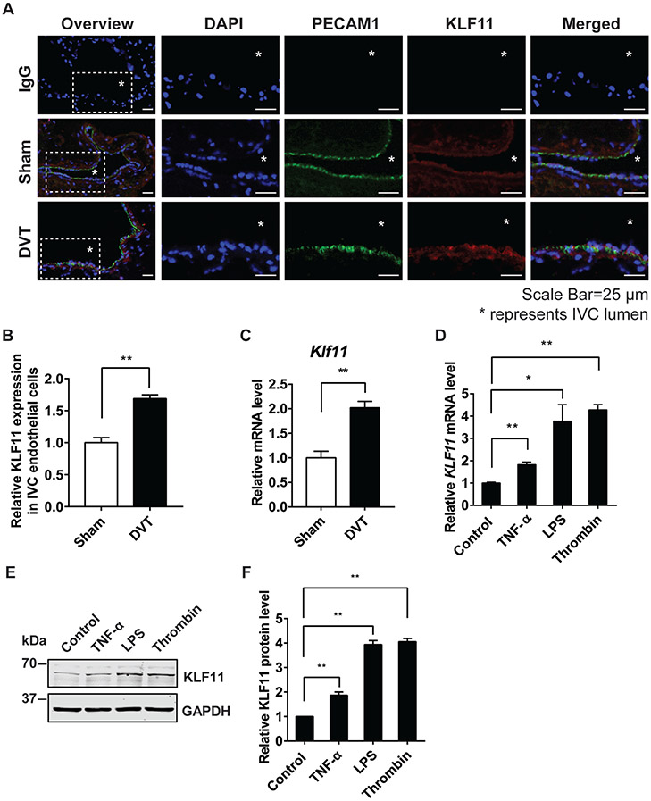
KLF11 expression is upregulated in endothelial cells under prothrombotic conditions. (A–C) In the stasis-induced deep vein thrombosis model (DVT), the inferior vena cava (IVC) from C57BL/6 male mice were either sham-operated or ligated for 48 hours to induce DVT (n = 3/group). The cryostat IVC sections were stained with antibodies against PECAM1 (DIA-310, Dianova, 1:100; Alexa 647, Jackson ImmunoResearch, 1:500, displayed in green) and KLF11 (X1710, Syd Labs, 1:50; Alexa 568, Jackson ImmunoResearch, 1:500, displayed in red). DAPI (4′,6-diamidino-2-phenylindole) stained for nuclear acid. Respective IgG staining was used as the negative control. Scale bars = 25 μm. The symbol “*” represents IVC lumen. The representative images are presented (A) and the expression of KLF11 in the endothelial cells of IVC was quantified by Image J (B). The vascular wall of IVC was isolated to measure Klf11 mRNA level (C). (D–F) Human umbilical vein endothelial cells (HUVECs) were treated with different stimuli for 4 hours to induce endothelial inflammation. The stimuli included: TNF-α (10 ng/mL), lipopolysaccharides (LPS, 100 ng/mL), and thrombin (5 U/mL). The mRNA level of KLF11 (D) was normalized to GAPDH and is presented relative to the control group. The representative Western blot (E) and quantification (F) of the KLF11 level are presented. N = 3/group. Data are presented as mean ± SEM. *p < 0.05, **p < 0.01 using unpaired Student t-test. IgG, immunoglobulin G; IVC, inferior vena cava; SEM, standard error of the mean; TNF-α, tumor necrosis factor-α.

