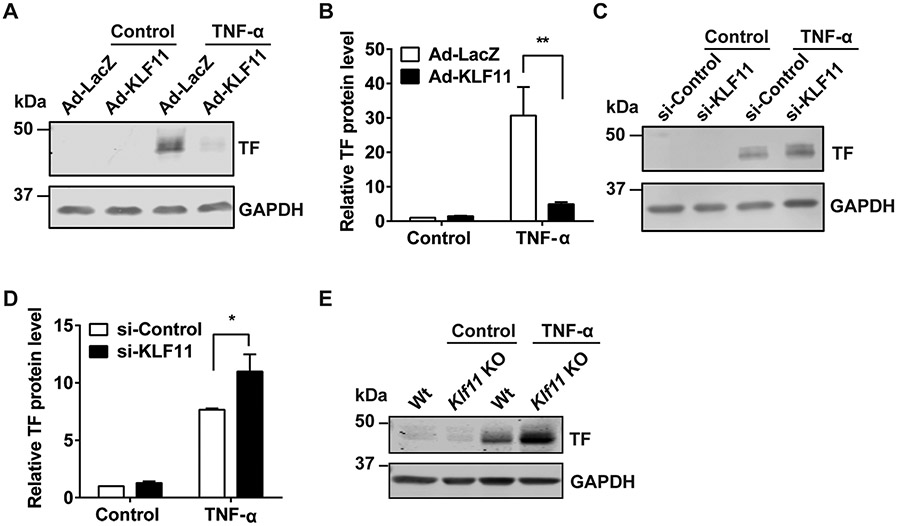
Fig. 3.
KLF11 negatively regulates TNF-α-induced tissue factor protein expression in endothelial cells. (A, B) Human umbilical vein endothelial cells (HUVECs) were infected with Ad-LacZ or Ad-KLF11 (10 MOI) for 48 hours, followed by TNF-α (10 ng/mL) stimulation for 4 hours. The representative Western blot of tissue factor (TF) protein level (A) and quantification (B) are presented. (C, D) HUVECs were transfected with si-Control or si-KLF11 (20 nM). Seventy-two hours after transfection, HUVECs were stimulated with TNF-α (10 ng/mL) for 4 hours. The representative Western blot of TF protein level (C) and quantification (D) are presented. Mouse aortic endothelial cells (MAECs) were isolated from wild-type (Wt) or Klf11 knockout (Klf11 KO) male mice (n = 3/group) and stimulated with TNF-α (2 ng/mL) for 4 hours. The representative Western blot of TF expression is presented (E). The mRNA level was normalized to GAPDH and is presented relative to the control group. N = 3/group. Data are presented as mean ± SEM. *p < 0.05, **p < 0.01 using two-way ANOVA followed by Bonferroni test. Antibodies: GAPDH (sc-32233, Santa Cruz Biotechnology, 1:1,000 dilution), tissue factor (sc-374441, Santa Cruz Biotechnology, 1:1,000 dilution). ANOVA, analysis of variance; SEM, standard error of the mean; TNF-α, tumor necrosis factor-α.

