Fig. 6.
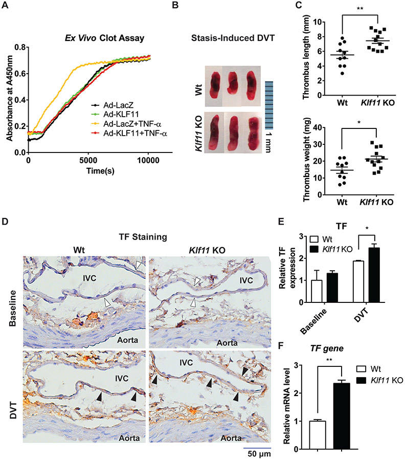
Endothelial KLF11 protects against thrombus formation. (A) Human umbilical vein endothelial cells (HUVECs) were infected with Ad-LacZ or Ad-KLF11 (10 MOI). Forty-eight hours after infection, HUVECs were stimulated with TNF-α (10 ng/mL) for 4 hours. Human plasma and calcium chloride were added, and the time of clot formation was measured. (B, C) The stasis-induced deep vein thrombosis (DVT) model was applied in 8–10 weeks old male wild-type (Wt, n = 10) or Klf11 KO (n = 12) mice by ligation of inferior vena cava (IVC) for 48 hours. Representative images of thrombus are shown. Bar = 1 mm (B). The length and weight of thromboses were measured (C). (D–F) The cross-sections of a combination of the abdominal aorta and IVC from Wt and Klf11 KO mice at baseline and in DVT condition (48-hour ligation) were prepared. (D) The tissue factor (TF) expression was determined by immunohistochemistry staining (filled arrowheads). The lumen of the aorta and IVC are labeled, and the endothelium of DVT is indicated with empty arrowheads. Bar = 50 μm. (E) The expression of TF was quantified by dividing the TF-positive endothelial area by the total endothelial area of IVC in ImageJ software. Data were analyzed with the Wt group at the baseline level set as 1. (F) The IVC from Wt and Klf11 KO mice were harvested after a 48-hour ligation procedure to detect the TF gene mRNA level (n = 4/group). *p < 0.05, **p < 0.01 using the unpaired Student’s t-test. TNF-α, tumor necrosis factor-α.

