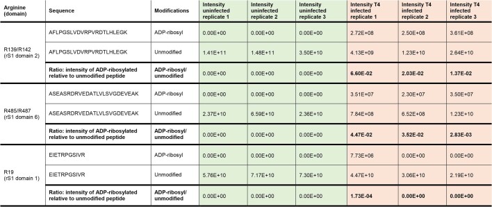
Extended Data Table 1.
ADP-ribosylation of endogenously His-tagged rS1 during T4 phage infection
MaxQuant intensities are presented for T4 phage-infected and uninfected samples in biologically independent triplicates (n = 3). R139/R142 located in rS1 domain 2 and R485/R487 in rS1 domain 6 appear as ADP-ribosylation sites on rS1 in vivo in all three replicates. The ratio comparing intensity of ADP-ribosylated and unmodified species of the same peptide is computed for each sample and peptide.

