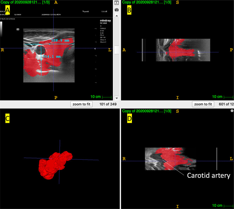
Fig. 5.
3D reconstruction of 1: ellipsoidal nodule in thyroid phantom, 2: spherical nodule in thyroid phantom, A original single frame scans from the US device, B Axial view, C Sagittal view, D Coronal view. Viewing options for the tissue surrounding the nodule were selected in the 3D view based on which view option highlighted the nodule the most clearly

