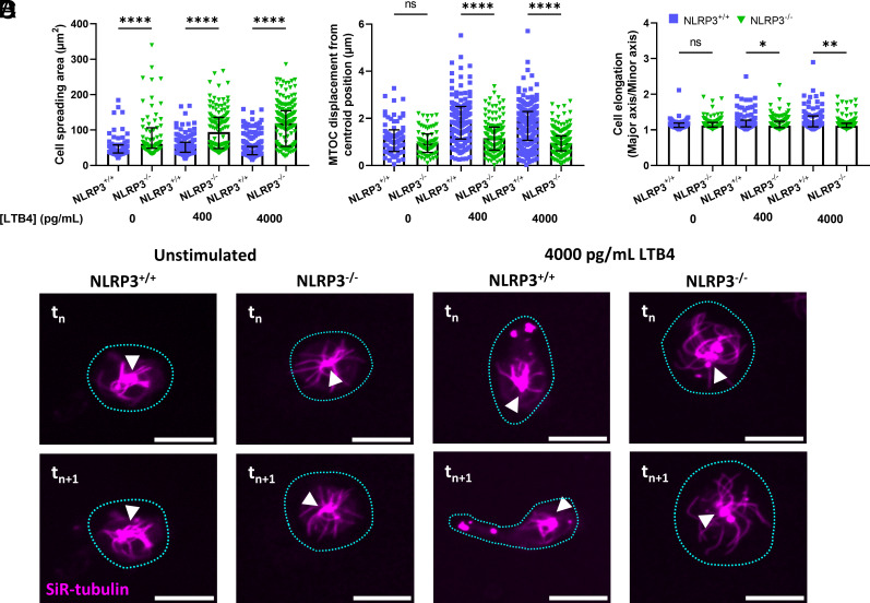
Fig. 3.
Deficiency in NLRP3 results in an inability of neutrophils to polarize in response to the presence of an LTB4 gradient. (A) Cellular spreading, as quantified by the surface area of the cell in the field of view, is significantly increased in the absence of NLRP3, both in the presence and absence of an LTB4 gradient. (B and C) quantification of neutrophil polarization through microtubule organizing center displacement (MTOC) and cellular elongation. (B) Displacement of the MTOC compared to the cell’s centroid position was taken as a measure for cellular polarization. NLRP3−/− neutrophils showed decreased capability for MTOC displacement away from the centroid position upon contact with a extracellular LTB4 gradient. (C) Cellular elongation, determined as the ratio of major and minor axis of the best fit ellipse on the cell. Again, a clear decrease in the propensity to elongate was observed in the NLRP3−/− neutrophils. (D) representative images of neutrophils, stained through SiR-tubulin, allowing for real-time visualization of the microtubule cytoskeleton. The shape of the cell is indicated through a blue dotted line, and the position of the MTOC is given by white arrowheads. A comparison between neutrophils at timepoint tn and 1 min later (tn+1) shows a clear polarization of NLRP3+/+ but not NLRP3−/− neutrophils, scale bar represents 10 µm. Kruskal–Wallis test was used for statistical analysis of two groups (P < 0.05 *, < 0.01 **, < 0.0001 ****, n = 3 to 4).

