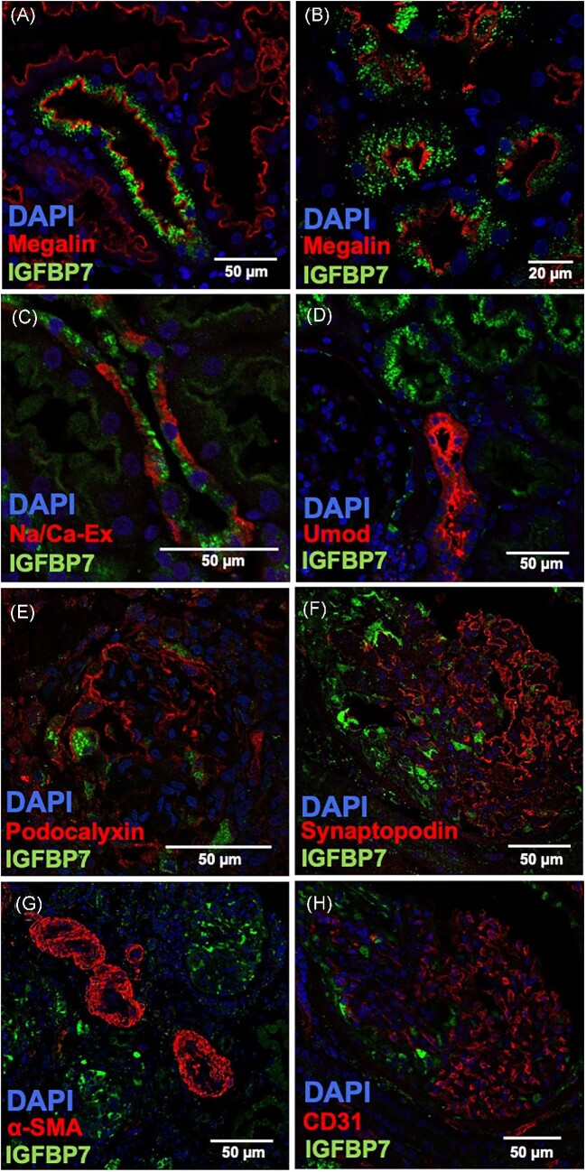
Figure 4:
Merged images of IGFBP7 and cell-specific markers are shown using immunofluorescence microscopy. IGFBP7 staining was combined with the marker megalin for (A, B) proximal tubules, (C) sodium/calcium exchanger (Na/Ca-Ex) for distal tubules, (D) uromodulin for the thick ascending limb, (E) podocalyxin and (F) synaptopodin for podocytes, (G) alpha smooth muscle actin (SMA) for vascular muscle cells and (H) CD31 for endothelial cells.

