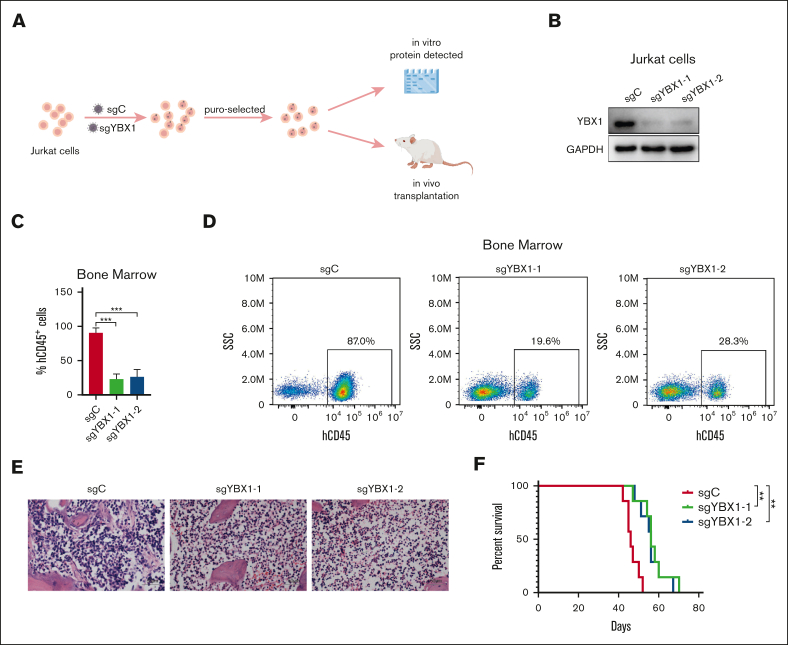
Figure 4.
YBX1 knockout decreases leukemia burden in the Jurkat xenograft model. (A) Graphical illustration of the Jurkat xenograft model. Jurkat cells were infected with lentiviruses carrying control (sgC) or YBX1 sgRNA (sgYBX1-1 or sgYBX1-2). The protein level of YBX1 was detected via western blot on day 5 after transfection and before transplantation. Approximately 4.5 × 10⁶ cells were injected into NCG mice intravenously, followed by the assessment of leukemia cell dissemination. (B) Protein analysis via western blot. (C) Flow cytometry analysis showing the percentage of hCD45+ cells in the BM on day 42 after transplantation. (D) Representative flow plot showing the frequency of hCD45+ leukemia cells in the BM. (E) Representative hematoxylin and eosin staining images of the BM from each group. (F) Survival curves are displayed using Kaplan-Meier plots (n = 7; long-rank test). ∗∗P < .01; ∗∗∗P < .001. The pattern diagram was drawn using Figdraw.

