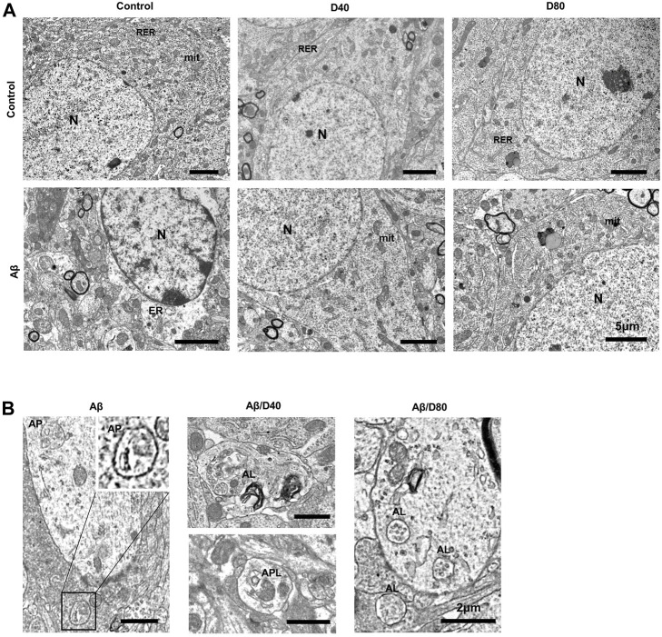
FIGURE 6.
The ultrastructural images of pyramidal neurons in the CA3 region in the hippocampus of C57B/6N mice on day 7 following Dex treatment, after i.c.v. injection of Aβ25-35 or saline (A) The ultrastructure of neurons in control showed well-organized nucleus (N), mitochondria (mit), and endoplasmic rough reticulum (RER). The D40 or D80 group had no differences compared to the control. The Aβ group showed rarefaction of cytosol, dilatation of the endoplasmic reticulum (ER) and nuclear chromatin agglutinates. The Aβ/D40 and Aβ/D80 groups were morphologically intact, similar to the control. (scale bar = 5 μm) (B) Magnified images of the autophagic vacuoles (AVs) after i.c.v. injection of Aβ25-35. In the Aβ group, A0056s observed were mostly double-membraned AVs, morphologically similar to autophagosomes (AP). In the Aβ/D40 and Aβ/D80 groups, single-membraned AVs (autolysosome, AL) were mainly observed and some autophagolysome (APL), autophagosomes combined with lysosomes, were also noticed, suggesting improvement of autophgic flux by Dex treatment. (scale bar = 2 μm).

