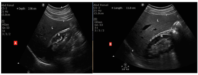
Figure 1.
Ultrasound images of two different cases: (A) a normal liver with similar brightness of the liver (L) and renal cortex (R), while (B) is a patient with grade 1 (mild) fatty changes showing the liver (L) is brighter than the renal cortex (R) but the wall of the intrahepatic vessels is still clearly seen.

