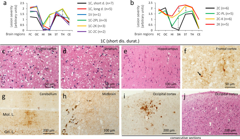
Fig. 2.
Lesion profiles of 9 sCJDMV histotypes, and histopathology of 1C. a sCJD MV1/MV1-2 can be divided into two major groups. The first one includes 1C, 1V and 1C-2C histotypes, characterized by the sparing of the substantia nigra (SN) and less severe pathological changes in thalamic (TH) nuclei; the second group encompasses 1C-2PL & 1C-2K showing more severe lesions of SN and TH, and accumulation of plaque-like PrP deposits (1C-2PL) and PrP plaques (1C-2K). b Lesion profiles of MV2-1 show three major groups: 2C with sparing of SN, 2C-PL with mild lesions of SN, and 2C-K & 2K with overall different lesion profiles, and marked hippocampal pathology; the 2C-K histotype shows C and K histotypic features in the proportion of ~ 50% each. Dashed double arrows point to differences in severity in key brain regions. c–e, j Hematoxylin–eosin (H&E). f–i PrP immunostaining. c–e Spongiform degeneration affecting the neocortex (c) and subcortical regions (d) regions, but not the hippocampus (e). f–h Diffuse PrP; arrow, f a larger PrP granule. Mol. L.: Molecular layer; Grl. L.: granular layer. i A focus of coarse PrP. j H&E preparation stained for PrP in i; antibody: 3F4; dis. durat.: disease duration

