Fig 1.
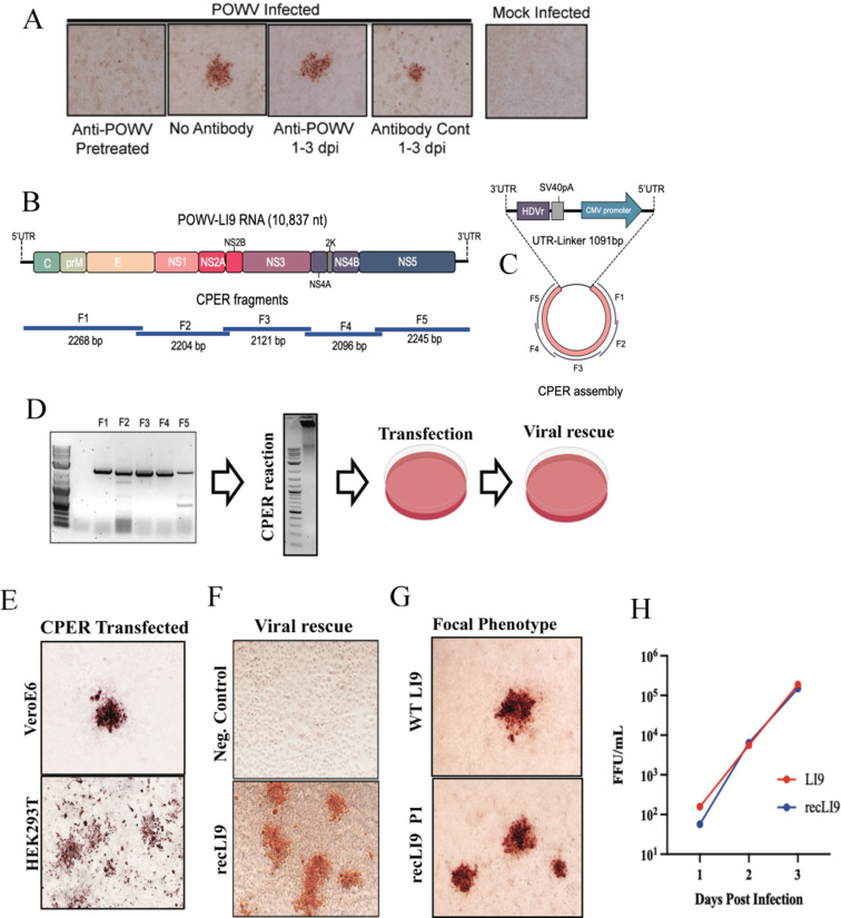
CPER generation and cell-to-cell spread of recombinant POWV strain LI9. (A) VeroE6 cells were infected with LI9 POWV at an MOI of 0.01 with or without anti-POWV HMAF (1:250) during adsorption. Cells were washed with PBS, and media were replaced with Dulbecco modified Eagle’s medium (DMEM) with or without anti-POWV HMAF (1:250) or control ascitic fluid 6 h post adsorption. Cells were methanol fixed 3 dpi and immunostained with anti-POWV HMAF (1:5,000) (20, 79, 80). (B) Schematic of the LI9 POWV genome and overlapping fragments amplified from LI9 cDNA. (C) CPER assembly schematic of F1-F5 fragments with a UTR-Linker fragment containing the last 26 nucleotides of the LI9 3′UTR, hepatitis delta virus ribozyme (HDVr), SV40 polyadenylation signal, a cytomegalovirus (CMV) promoter, and 33 nucleotides of the LI9 5′UTR. (D) Agarose gel electrophoresis of PCR-amplified fragments (F1–F5) showing a representative image of three experimental repeats. F1–F5 were combined in equal molar amounts with the UTR-Linker in a CPER. A representative agarose gel of CPER product. Resultant LI9 CPER products were transfected into HEK293T or VeroE6 cells, and supernatants were subsequently used to infect VeroE6 cells and rescue infectious recLI9 viruses. (E) Immunostaining CPER-transfected VeroE6 or HEK293T cells (7 or 3 dpt, respectively) display focal cell-to-cell spread foci morphologies. (F) Infectious recLI9 virus rescued from CPER transfected cells and grown in HEK293T cells vs CPER controls amplified without the UTR-linker fragment. (G) Comparison of WT LI9 and recLI9 focal cell-to-cell spread phenotypes in immunostained VeroE6 cells. (H) Growth kinetics of WT LI9 (red) and recLI9 (blue) POWVs 1–3 dpi in VeroE6 cells (MOI 1). Analysis repeated at >3 times.

