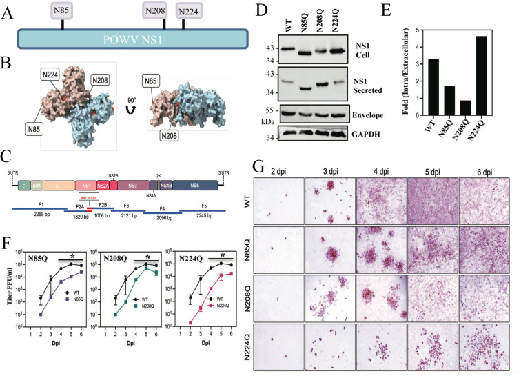
Fig 2.
CPER POWV NS1 glycosylation mutants have impaired replication kinetics. (A) Schematic of N-linked glycosylation sites within POWV NS1 proteins. (B) Alphafold2 model of dimeric POWV NS1 protein showing pink or blue monomers and the localization of putative N-linked glycosylation sites (N85, N208, N224) in red (84). (C) Schematic strategy for CPER-generating N-linked glycosylation site (NxT) mutants using overlapping fragments to introduce N to Q codon changes. Fragment 2 was split into subfragments 2A and 2B with overlapping regions incorporating mutations. (D) VeroE6 cells were infected with WT LI9, recLI9-NS1N85Q, recLI9-NS1N208Q, and recLI9-NS1N224Q mutant viruses (MOI 1), and 7 dpi cell lysates and supernatants were analyzed by Western blot using antibodies POWV-NS1 (mAb), anti-POWV HMAF, or GAPDH. (E) Ratios of intracellular vs extracellular NS1 levels from representative Western blot (2D) of rec-LI9-NS1 glycosylation mutants were performed >2 times. (F) Growth kinetics of WT LI9 (black), CPER-generated NS1 glycosylation mutants recLI9-NS1N85Q (purple), recLI9-NS1N208Q (green), and recLI9-NS1N224Q (pink) 1–6 dpi of VeroE6 cells (MOI 0.1). *Statistically significant by two-way ANOVA with Tukey’s post-test (P < 0.05). (G) Kinetic comparison of focal cell-to-cell spread by WT LI9 and recLI9-NS1 glycosylation mutants 1–6 dpi in VeroE6 cells (MOI 0.1) by anti-POWV (HMAF) immunoperoxidase staining of POWV-infected cells.

