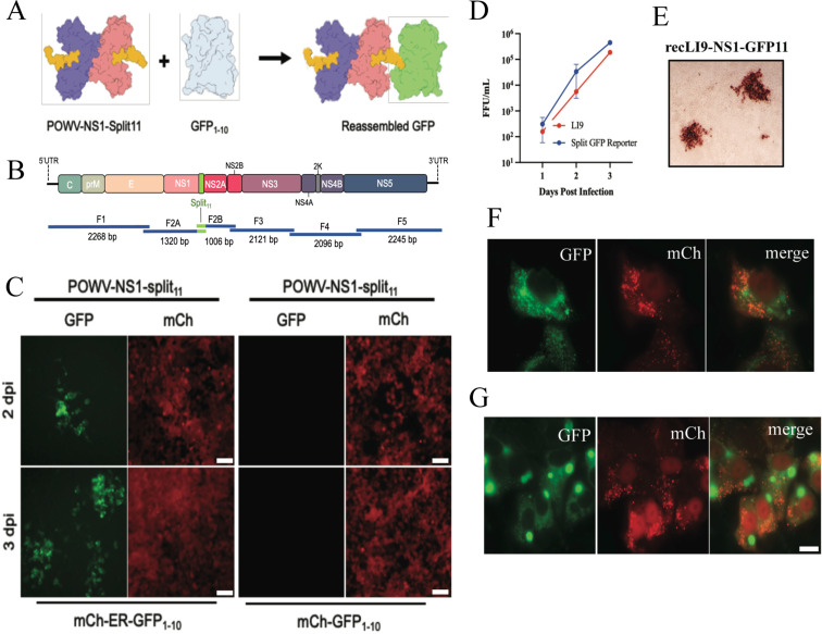
Fig 5.
Generation of a split GFP POWV reporter virus. (A) Schematic of the split GFP fluorescence reporter system directed by the recLI9-NS1-GFP11 virus. Individual NS1 monomers (blue and pink) with a 16 residue GFP11 tag added to the NS1 C-terminus (yellow) with co-expressed, nonfluorescent, GFP1–10 (gray). The recLI9-NS1-GFP11 expression of NS1-GFP11 protein reconstitutes GFP fluorescence in cells co-expressing GFP1–10. (B) CPER strategy for POWV-NS1-split11 generation. F2 was split into subfragments F2A and F2B with primer-directed incorporation of GFP11 sequences. (C) Retrovirus-transduced HEK293T cells constitutively expressing mCherry (cytoplasm) and either ER-localized ER-GFP1–10 (mCh-ER-GFP1–10) or cytoplasm-localized GFP1–10 (mCh-GFP1–10) were infected with recLI9-NS1-GFP11. Live imaging captured 2–3 dpi shows foci of GFP fluorescence in LI9-NS1-GFP11-infected cells expressing ER translocated GFP1–10 but not cytoplasmically expressed GFP1–10. (D) Growth kinetics of WT LI9 (red) and recLI9-NS1-GFP11 (blue) 1–3 dpi in VeroE6 cells (MOI 1). (E) Immunostaining of LI9-NS1-GFP11 infected VeroE6 cell foci 3 dpi with anti-POWV HMAF. (F and G) Retrovirus-transduced VeroE6 cells expressing mCherry-ER-GFP1–10 were LI9-NS1-GFP11 infected, and 6 dpi cells paraformaldehyde fixed. Representative images show perinuclear NS1-GFP fluorescence (F) and the discrete accumulation of NS1-GFP in large intracellular vesicles 6 dpi (G). Bars represent 10 µm.

