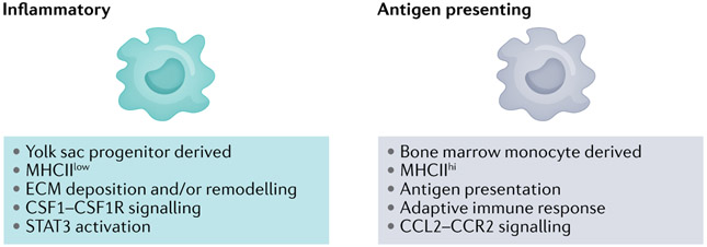
Fig. 3 ∣. Macrophage lineages in pancreatic cancer.
Macrophages found in tissues can be tissue resident, yolk sac derived or differentiate from bone marrow-derived inflammatory monocytes. Single-cell transcriptomics identified three macrophage subclusters in the normal pancreatic gland, ultimately resolving into two populations – inflammatory-like and major histocompatibility complex II (MHCII)-rich – in both pre-invasive disease and invasive disease that blended and divided the combined transcriptional repertoires between them (that is, the two classes that emerged in invasive cancer were distinct from the three that preceded them in the normal gland)51. In the normal gland, the three cell populations appear to retain some degree of fluidity and do not fully adopt their ultimate phenotypes until later in disease progression. The inflammatory signature also appeared to increase with disease progression51, and a recent study has identified the yolk sac-derived macrophages to be tumour promoting and, therefore, more like the so-called M2 phenotype154. CCL2, CC-chemokine ligand 2; CCR2, CC-chemokine receptor 2; CSF1, macrophage colony-stimulating factor 1; CSF1R, macrophage colony-stimulating factor 1 receptor; ECM, extracellular matrix; STAT3, signal transducer and activator of transcription 3.

