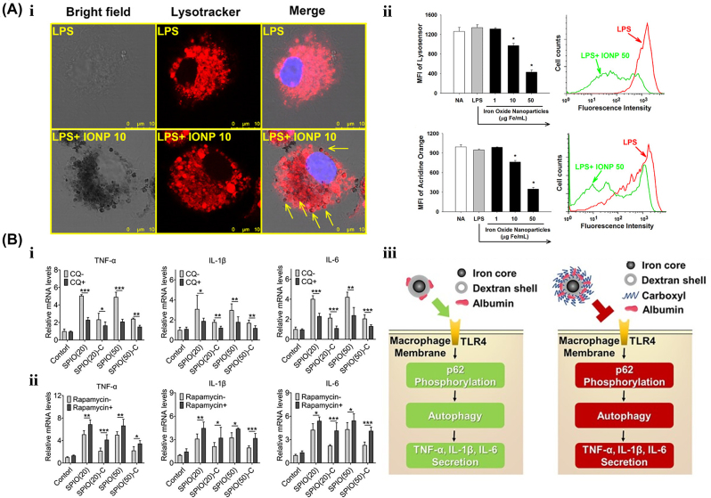
Figure 7:
The Lysosomal-autophagy modulation effect by iron-based nanoparticles. A. After pre-treatment with IONPs (1–50 μg Fe/mL), (i) the morphology of lysosomes with Lysotracker (red) staining, (ii) lysosome acidity measured by lysosome sensor and (iii) cell permeability with acridine orange staining in microglia stimulated by LPS. IONPs treated cells had scattered dark brown dots. The arrows represent the colocalization of dark brown dots and lysosomes. Reproduced with permission [111]. Copyright 2013, Springer Nature. B. The relative mRNA levels of TNF-α, IL-1β, and IL-6 in SPIO-treated RAW264.7 cells (i) with autophagy inhibitor CQ and (ii) autophagy enhancer rapamycin, respectively. (iii) Schematic diagram of carboxyl-modified SPIONs inhibited macrophage autophagy and inflammatory cytokine secretion. Reproduced with permission [96].

