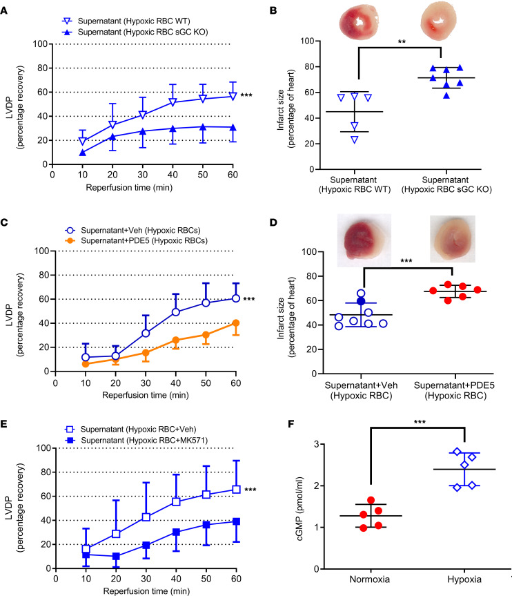
Figure 2. Hypoxia-induced release of cardioprotective cGMP from RBCs.
(A) Percentage recovery of LVDP and (B) infarct size following administration of supernatant from hypoxic RBCs from WT mice (n = 5) or sGC KO mice (n = 7). (C) Recovery of LVDP and (D) infarct size following administration of supernatant from hypoxic RBCs incubated with vehicle(veh; n = 8) or recombinant PDE5 (n = 6). (E) Recovery of LVDP following administration of supernatant from hypoxic RBCs incubated with vehicle (n = 11) or the cGMP transport inhibitor MK571 (n = 5). (F) cGMP in supernatant of RBCs exposed to normoxia and 1 hour of hypoxia. Data are presented as mean ± SD. Data points in the D Supernatant Hypoxic RBC+Veh group are also shown as open and filled circles in 1B and open circles in the Figure 4D Supernatant Hypoxic RBC group. The same overlapping data points in the Supernatant Hypoxic RBC group in C are also included in Figures 1A and 4C in the Supernatant Hypoxic RBC group. **P < 0.01 and ***P < 0.001 denote significant differences between groups using 2-way ANOVA followed by Tukey’s multiple comparison test in A, C and E, Mann-Whitney test in B, and an unpaired t test in D and F.

