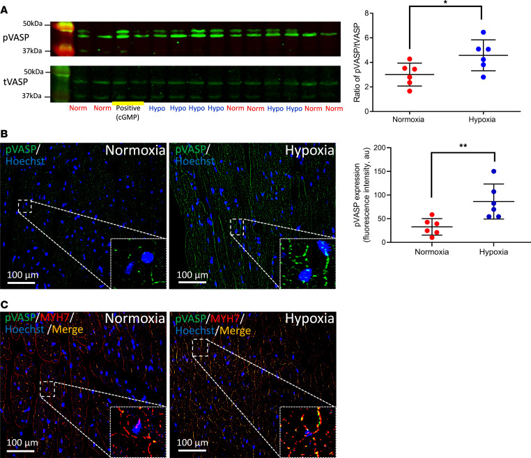
Figure 8. Increased expression of phosphorylated VASP in cardiomyocytes by hypoxic RBCs.
(A) Expression of phosphorylated and total VASP (pVASP and tVASP, respectively) using Western blot in cardiac tissue following administration of supernatant from hypoxic (Hypo; n = 6) or normoxic (Norm; n = 6) RBCs or cGMP (0.1 mmol/L, positive control) to Langendorff-perfused mouse hearts subjected to 5 minutes ischemia and 1 minute reperfusion. (B and C) Immunofluorescence showing expression of pVASP following administration of supernatant from RBCs exposed to normoxia (n = 6 for both B and C) or hypoxia (n = 6 for both B and C). Immunoreactivity of pVASP was visualized using Alex Fluor 488 antibody (green in B) and the cardiomyocyte specific marker myosin heavy chain 7 (MYH7; red in C). Merged staining is yellow. Nuclei were stained with Hoechst (blue). Quantitative data in A and B are presented as mean ± SD. *P < 0.05 and **P < 0.01 denote significant differences by unpaired t test.

