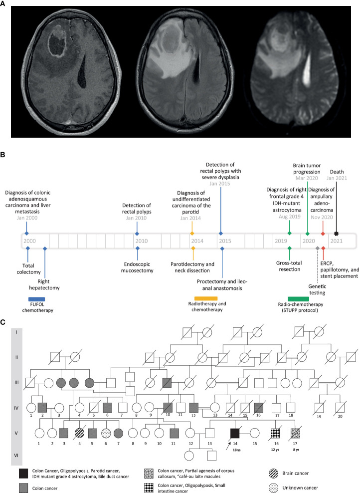
Figure 1.
(A) Preoperative cranial MRI of the proband (from left to right: axial T1-weighted with gadolinium injection, axial T2 FLAIR-weighted, axial apparent diffusion coefficient). There is a solid intra-axial mass within the right frontal lobe with marked ring-like enhancement and an internal necrotic area. The enhancing component of the lesion shows diffusion restriction. (B) Timeline showing the clinical history of the patient. (C) Family pedigree. The black arrow indicates the proband case. The age of the first tumor is given below the proband and the two affected brothers.

