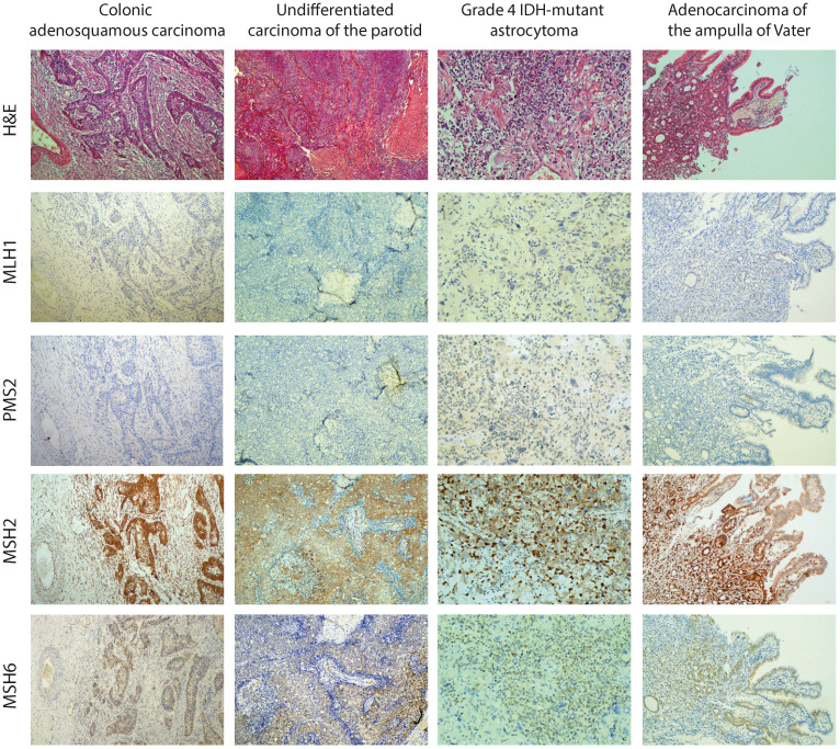
Figure 3.
Histology (hematoxylin and eosin (H&E)) and immunohistochemical staining for all four MMR proteins (MLH1, PMS2, MSH2, and MSH6) in all tumor specimens of the proband (colonic and parotid tumor at 40× magnification, brain tumor and pancreatic tumor at 100× magnification). As the representative images show, immunostaining for MLH1 and PMS2 revealed a loss of expression in neoplastic cells and surrounding normal cells.

