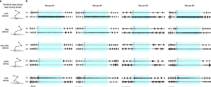
Extended Data Fig. 4. Stimulation of Chx10-PPN neurons at different phases of the step cycle leads to unique arrest patterns that are reproducible across animals.
Chronic hindlimb EMG recordings during locomotion in a linear corridor paired with optogenetic stimulation of Chx10-PPN neurons. a, Hindlimb step phase kept during the optogenetically evoked locomotor arrest depicted by stick diagrams. The elliptical shade below the foot indicates the step cycle phase (gray, stance; red, swing). b, Matrix showing examples of muscle activity patterns when the same animal (first and second columns, mouse #1) or different animals (third and fourth columns, mice #2 and #3) were arrested in the same phase of the step cycle (across rows) and at different phases of the step cycle (across columns). The step phase for each row corresponds to the diagram in a. Every pair of traces in the same row shows the muscle activity of the Tibialis anterior (TA, upper trace) and the Soleus (Sol, bottom trace) during the evoked arrest. The arrest evoked by Chx10-PPN neuron activation at any given phase of the step cycle leads to a similar posture and EMG pattern in TA and Sol both in the same animal and across animals. Purple arrowheads and lines mark the moment of movement arrest for each hindlimb. Blue shades delimit light stimulus duration (2 s —mice #1 and #3— or 1 s —mouse #2— at 40 Hz).

