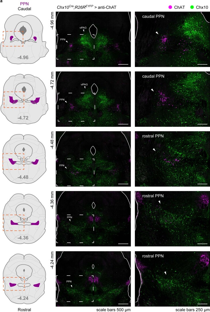
Extended Data Fig. 1. Mutually exclusive distribution of Chx10+ and ChAT+ neurons along the rostro-caudal axis of the PPN.
a, Left, coronal plane schematics highlighting the PPN (magenta) at different rostro-caudal levels. Right, confocal photomicrographs showing the distribution of Chx10+ (green, Chx10Cre; R26EYFP reporter mouse) and ChAT+ (magenta, antibody) neurons at the corresponding rostro-caudal levels of the schematics to the left. The pictures at the right-most column are magnified images corresponding to the area delineated by dashed lines in the pictures to the left. The same area is also delineated in the schematics with orange dashed lines. White arrowheads in the magnified pictures point to the PPN. Chx10+ neurons are sparse within the caudal half of the PPN while their density increases in the rostral half of the PPN, following an inverted pattern compared to ChAT+ neurons within the nucleus. There is no overlap in the cellular expression of ChAT and Chx10 at any rostro-caudal level of the PPN. Chx10+ neurons are also found in the vlPAG where they are enriched at the coronal levels corresponding to the caudal half of the PPN (see also Extended Data Fig. 8).

