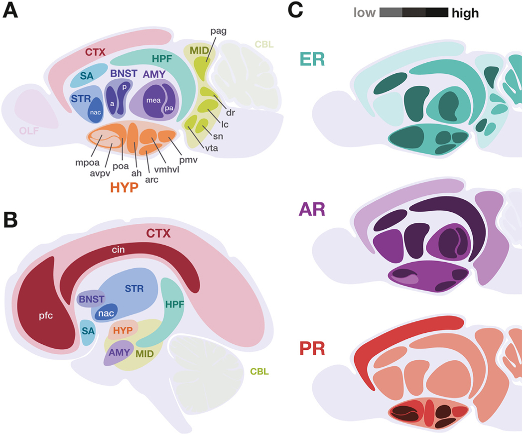
Figure 2.
Key neuroanatomical regions to consider in gender-affirming hormone therapy studies in both rodent animal models and humans. Certain brain regions are especially relevant to the study of gender-affirming hormone therapies, indicated in (A) murine and (B) human central nervous systems. Broad anatomical regions are indicated by uppercase abbreviations, with key subregions indicated in lowercase. (C) Schematic summarizing the relative expression levels of canonical nuclear hormone receptors in the murine brain, with darker shades indicating higher expression. Data represented includes both immunolocalization of receptor protein and receptor messenger RNA in adult rodent models. This summary does not include membrane or other hormone receptors (Table 1) and does not account for known isoform, developmental, and sex variability. [Visualizations based on data presented in (67,71,90–92)]. a, anterior; ah, anterior hypothalamus; AMY, amygdala; AR, androgen receptor; arc, arcuate nucleus; avpv, anteroventral periventricular nucleus; BNST, bed nucleus of the stria terminalis; CBL, cerebellum; cin, cingulate; CTX, cortex; dr, dorsal raphe; ER, estrogen receptor; HPF, hippocampal formation; HYP, hypothalamus; lc, locus coeruleus; mea, medial amygdala; MID, midbrain neuromodulatory centers; mpoa, medial preoptic area; nac, nucleus accumbens; OLF, olfactory bulb; p, posterior; pa, posterior amygdala; pag, periaqueductal gray; pfc, prefrontal cortex; pmv, ventral premammillary nucleus; poa, preoptic area; PR, progesterone receptor; SA, septal area, includes medial and lateral septal nuclei; sn, substantia nigra; STR, striatum; vmhvl, ventromedial hypothalamus ventrolateral division; vta, ventral tegmental area.

