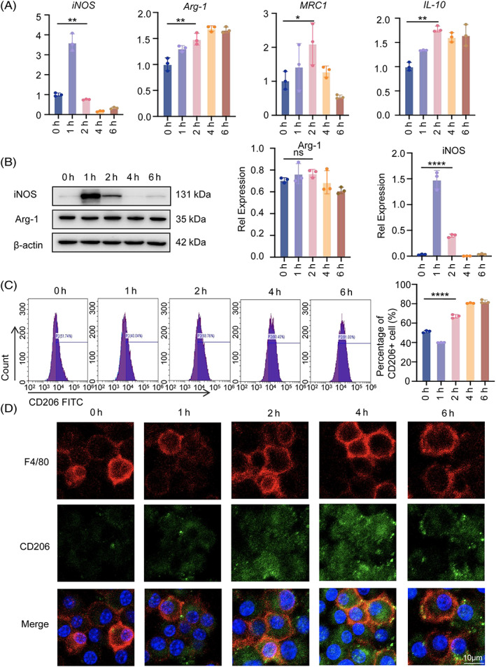
FIGURE 1.

Mechanical tension induces macrophage polarization. (A) Real‐time RT‐PCR was used to assess the mRNA levels of iNOS, Arg‐1, MRC1 and IL‐10 in RAW264.7 cells as a result of the 0–6 h of tension. (B) Western blot was used to identify the protein expression of iNOS and Arg‐1 in RAW264.7 cells under stress for 0–6 h. GAPDH was utilized as the standard to quantify iNOS and Arg‐1. (C) The expression of CD206 in RAW264.7 cells under tension was detected by flow cytometry, and the statistical results were displayed. (D) M2 macrophages in RAW264.7 cells under the tension of 0–6 h were visualized using immunofluorescence staining of F4/80 (red) and CD206 (green). The cell nuclei were detected with DAPI. Scale bar: 10 μm. Data represent the mean ± SD. *p < 0.05, **p < 0.01, ***p < 0.001.
