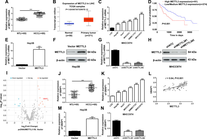
Fig. 1.
Upregulated METTL3 increases GBAP1 expression in HCC. A RT-qPCR analysis was applied to explore METTL3 expression in HCC tissues (n = 85) and adjacent non-tumor tissues (n = 85) (mean ± SD; n = 3). ***P < 0.001 (Student’s t test). B Data from TCGA showed that METTL3 expression was significantly increased in HCC tissues (n = 371) compared to normal tissues (n = 50) (https://ualcan.path.uab.edu/index.html) (Student’s t test). C RT-qPCR analysis was applied to explore METTL3 expression in HCC cell lines and normal hepatic cell LO2. ***P < 0.001 versus LO2 (two-way ANOVA). D Data from UALCAN platform showed that HCC patients with higher METTL3 expression had worse prognosis than that with lower METTL3 (https://ualcan.path.uab.edu). E and F RT-qPCR analysis and Western blot were applied to verify the overexpressing efficiency of pcDNA/METTL3 in Hep3B cells. ***P < 0.001 (Student’s t test). G and H RT-qPCR analysis and Western blot were applied to verify the knockdown efficiency of METTL3 shRNA#1 and shRNA#2 in MHCC97H cells. ***P < 0.001 versus shNTC (two-way ANOVA). I RNA-seq was conducted in METTL3 overexpressing and the control sunclones of Hep3B. Scatter diagram was applied to show the differentially expressed genes which were regulated by METTL3 in Hep3B cells. J RT-qPCR analysis was applied to explore GBAP1 expression in HCC tissues (n = 85) and adjacent non-tumor tissues (n = 85). ***P < 0.001 (Student’s t test). K RT-qPCR analysis was applied to explore GBAP1 expression in HCC cell lines and normal hepatic cell LO2. ***P < 0.001 versus LO2 (two-way ANOVA). L Pearson correlation analysis showed that there existed a positive correlation between METTL3 and GBAP1 in HCC tissues (n = 85). M and N RT-qPCR analysis showed that GBAP1 was significantly increased by pcDNA/METTL3 in Hep3B cells, while GBAP1 was significantly decreased by METTL3 shRNAs. ***P < 0.001 (Student’s t test). ***P < 0.001 versus shNTC (two-way ANOVA)

