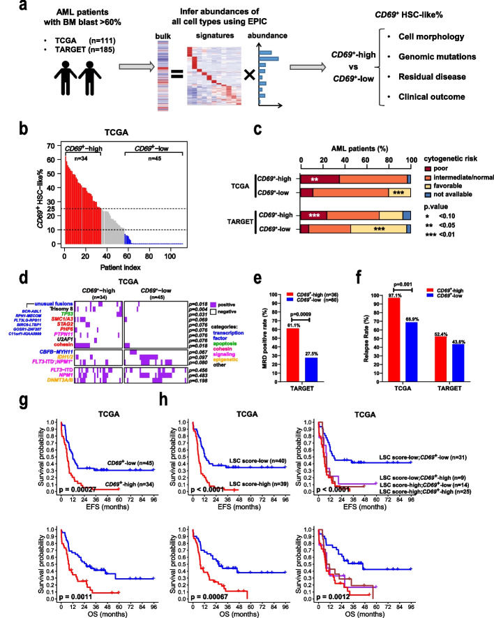
Fig. 7.
Clinical and genomic features of AML patients with different CD69+ HSC-like cell proportions. a Schematic workflow illustrating the exploration of the clinical relevance of the CD69+ HSC-like subpopulation in two large public AML cohorts. b Estimated proportions of CD69+ HSC-like subpopulation (CD69+ HSC-like%) in pre-therapy samples from TCGA-AML patients. Patients were grouped into CD69+-high (red), CD69+-middle (grey), and CD69+-low (blue) according to CD69+ HSC-like%, with dashed black lines indicating the cutoffs. c Histogram showing the percentages of patients with different cytogenetics-based prognostic risk categories in each group. d Heatmap showing the presence of genomic alterations in samples from TCGA-AML patients. Genomic alterations (rows) are colored according to the biological functions of their corresponding genes. The cohesin term includes mutations of the core complex subunits STAG2, RAD21, SMC1A/3/5, or the modulator PDS5B. FLT3-ITD−;NPM1+ represents mutated NPM1 without FLT3-internal tandem duplication (ITD). Unusual fusions are indicated. e Flow cytometry-based measurable residual disease (MRD) positive rates in the TARGET-AML patients at the end of the first cycle of chemotherapy regimen. A patient was defined as MRD-positive if the MRD level was equal to or greater than 0.1%. f Relapse rates in two groups of AML patients from each cohort. g Kaplan–Meier curves showing the event-free survival (EFS) and overall survival (OS) of TCGA-AML patients stratified by CD69+ HSC-like%. h Kaplan–Meier curves showing the survivals of TCGA-AML patients stratified by LSC score alone or combined with CD69+ HSC-like%. All p values in panels c, d, e, and f were calculated using Fisher’s test. All p values in panels g and h were calculated using the log-rank test

