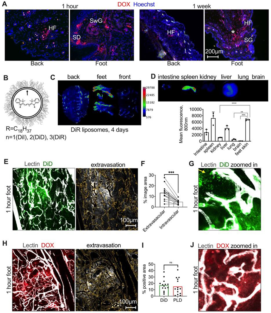
Fig. 1. Extravasation of liposomes in different skin areas.

A) Representative images of accumulation of PEGylated liposomal doxorubicin (PLD, LipoDox®) in different areas of the skin. HF, hair follicle; SD, sweat duct; SwG, sweat gland; SG, sebaceous gland; asterisk shows deep loose dermis; B) HSPC/Chol/DSPE-PEG2000 liposomes were labeled with indocarbocyanine dyes; C) pseudocolored ex vivo images 4 days after i.v. injection of DiR labeled liposomes show enhanced accumulation in feet and in the areas subject to mechanical stress/pressure in the back and front skin (arrows); D) Accumulation in the foot skin is higher than in main organs except spleen (n=3 mice, 2 way ANOVA); E) mice were injected i.v with HSPC/Chol/DSPE-PEG2000 liposomes labeled with DiD, and then FITC-lectin to label blood vessels. Mice were extensively perfused with PBS to remove the blood and remaining liposomes in all experiments. Representative confocal images of excised foot skin 1h post-injection show intense “bursts” of extravasation. Image on the right shows the extravascular fluorescence after blood vessel thresholding (outlined as yellow trace); F) quantification of intravascular and extravascular fluorescence in multiple areas of foot skin (n=13 images, 2 feet, paired t-test); G) “zoomed in” area of the extravasation “burst” with discrete particles (arrow) and diffuse fluorescence; H) representative confocal images of excised foot skin 1h post-injection of PLD show similar burst-like extravasation (center). Image on the right shows the extravascular fluorescence after blood vessel thresholding (outlined as yellow trace); I) extravasation of doxorubicin fluorescence is comparable to the extravasation of PEGylated DiD liposomes (n=2 feet per group, repeated twice, two-sided t-test); J) representative “zoomed in” area showing doxorubicin extravasation.
