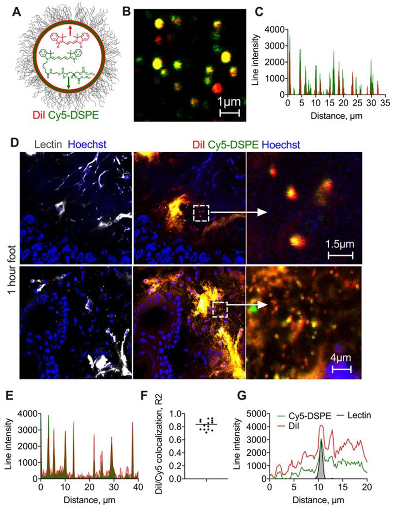
Fig. 3. Double labeled liposomes extravasate as intact particles and as diffusing lipids.

A) EggPC/DSPE-PEG2000 liposomes were labeled with lipophilic membrane dyes DiI and Cy5-DSPE; high magnification image (B) and line profile (C) show colocalization. Size is overestimated in the fluorescence images but is less than 1μm for most of the liposomes; D) representative confocal images of foot skin histological sections (dermis) 1h post-injection of liposomes (2 different areas). “Zoomed in” images show dual labeled extravasated particles, likely intact liposomes. Size of extravasated particles (albeit overestimated in fluorescence images) is less than 1μm; Diffusion and spreading of both dyes away from blood vessels is also seen. Repeated in 2 mice; E) line profile drawn across multiple extravascular particles shows colocalization of both dyes; F) Person colocalization coefficient for multiple particles; G) representative line profile across lectin-stained endothelium shows spreading of both dyes outwards.
