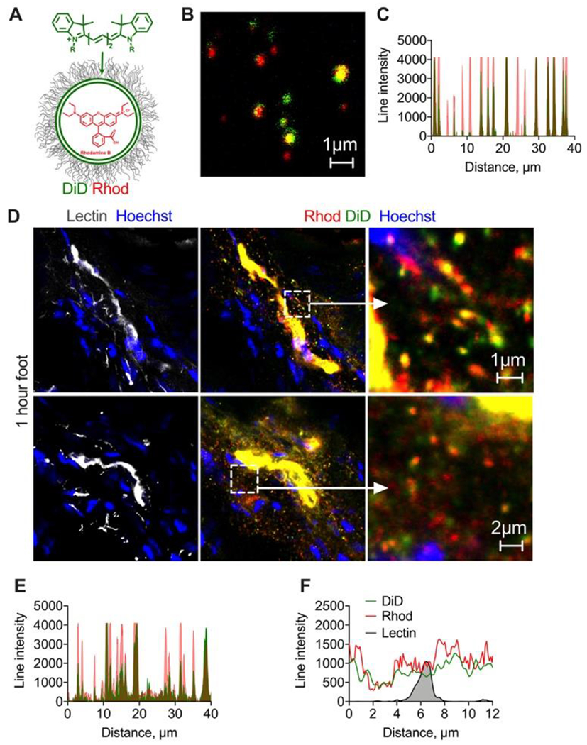
Fig. 4. Membrane- and internally-labeled liposomes extravasate as intact particles and as individual components.

A) EPC/DSPE-PEG2000 liposomes were labeled with lipophilic membrane dyes DiD and internally with rhodamine B; high magnification image (B) and line profile (C) show colocalization of both labels for the majority of liposomes, although the labels’ ratio is variable. Size is overestimated in fluorescence images but is less than 1μm; D) representative confocal images of sections of foot skin (dermis) 1h post-injection of dual labeled liposomes (2 different areas). Cropped images show extravasated liposomes. Diameter of extravasated particles (overestimated in fluorescence images) is less than 1μm. Diffusion of both dyes from the blood vessels can be observed. Repeated in 2 mice; E) line profile drawn through multiple extravasated particles shows colocalization of both dyes; F) representative line profile drawn across lectin+ endothelium shows spreading of both dyes. Experiment was repeated twice.
