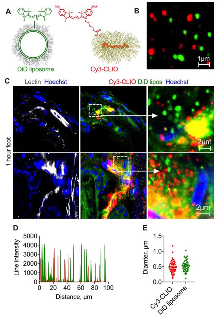
Fig. 5. Solid nanoparticles extravasate only as intact particles.

A-B) EPC/DSPE-PEG2000 liposomes labeled with DiD and similarly-sized CLIO NWs labeled with sulfo Cy3 were injected in the same mouse; C) representative confocal images of histological sections of foot skin (dermis) 1h post-injection of liposomes and nanoparticles (2 different areas) show that liposomes extravasated both as intact particles and as migrating DiD, whereas Cy3 extravasated only as intact particles. Repeated in 2 mice; D) line profile drawn though multiple extravascular particles shows almost no colocalization between Cy3-CLIO and DiD liposomes; E) diameter of extravasated liposomes and nanoparticles (overestimated in fluorescence images) is similar and mostly below 1μm.
