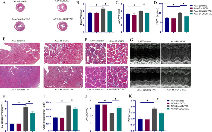
Fig. 2.
Inhibition of UGCG expression in the heart relieved pathological remodeling of myocardial hypertrophy. A Masson staining showed the overall degree of myocardial hypertrophy and fibrosis. B–D Statistical charts of HW/BW, LW/BW, and HW/TL in different groups. E, H Masson staining showed fibrosis at the same site in the hearts of different groups of mice and corresponding statistical analysis histogram. F, I The representative pictures of the size of the cross-sectional area of cardiomyocytes in the heart and corresponding statistical analysis histogram. G, J, K Echocardiogram and index results of each group. N = 5, *p < 0.05. AAV-Scramble: negative control of AAV-Sh-UGCG; AAV: Adeno-associated virus; Sh: Small hairpin; LVIDd: Left ventricle internal dimension diastole; LVPWd: Left ventricle posterior wall dimension

