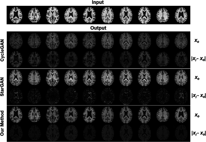
FIGURE 11.

Visual comparison across three methods (CycleGAN, StarGAN, our style‐encoding generative adversarial network [GAN]) of image harmonization using 10 images from subjects within the ADNI dataset, harmonized to a single subject within the UK Biobank (UKBB) dataset. For each method, the first row is the harmonized images translated from the input, and the second row is the absolute difference between the input and the harmonized images. Anatomical structure is not preserved in CycleGAN or StarGAN as specifically evident by the alterations within the ventricles and other contrast differences around tissue boundaries.
