Figure 5.
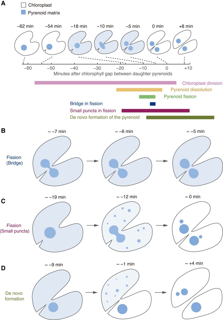
The Chlamydomonas pyrenoid exhibits liquid-like behavior during cell divisions. A) Diagram depicting the timeline and morphology of a typical cell division with pyrenoid fission in Chlamydomonas (adapted from Freeman Rosenzweig et al. 2017). The time point t = 0 is the moment the chloroplast division furrow passes between the daughter pyrenoids. A portion of the pyrenoid matrix disperses into the chloroplast stroma during the division of the pyrenoid. The approximate timing and duration of key events are shown below the timeline. B) Diagram depicting the “bridge” of matrix during pyrenoid fission. C) Diagram depicting the transient appearance of small puncta of pyrenoid matrix throughout the stroma during dispersal of the matrix in some dividing cells. D) Diagram depicting the de novo formation of a daughter pyrenoid when pyrenoid fission fails. The lower daughter cell inherits the entire pyrenoid of the mother cell. The upper cell shows de novo pyrenoid formation with the appearance of one or more fluorescent puncta growing or coalescing into one pyrenoid (observed in wild-type cells expressing either EPYC1-Venus or Rubisco-Venus).

