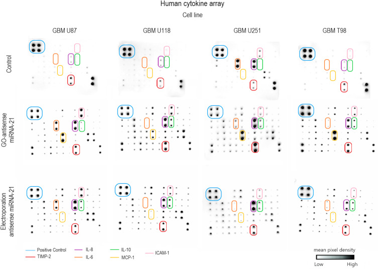
Figure 5.
Cytokine antibody array. Control – non-treated glioblastoma cell line. Glioblastoma cell line: U87, U118, U251, and T98. The arrows point to significantly changed pixel densities of positive dots of protein: interleukin 6 (IL-6), interleukin 8 (IL-8), interleukin 10 (IL-10), intercellular adhesion molecule 1 (ICAM-1), monocyte chemoattractant protein-1 (MCP-1), and metallopeptidase inhibitor 2 (TIMP-2).

